
 Descargar número completo
 Download full issue
CITE THIS WORK
Alonso Belmonte C, Parra López B, Sánchez Sánchez MI, Jiménez Pérez M, Bravo Aranda AM. Hepatocellular carcinoma in a patient with acute intermittent porphyria: importance of digestive follow-up. RAPD 2026;49(4):138-141. DOI: 10.37352/2026494.3
Introduction
Porphyrias are a heterogeneous group of rare metabolic disorders—either hereditary or acquired—caused by abnormalities in the activity of enzymes involved in heme biosynthesis, leading to the accumulation of toxic precursors with systemic effects [1]. Depending on the location of the enzymatic defect, they are classified as hepatic or erythropoietic porphyrias, and depending on their clinical presentation, as acute or cutaneous forms.
Acute intermittent porphyria (AIP) is the most common form of hereditary acute porphyrias. It is caused by a deficiency of the enzyme porphobilinogen deaminase, leading to the accumulation of delta-aminolevulinic acid (ALA) and porphobilinogen (PBG), both of which have neurotoxic effects[1],[2]. It is inherited in an autosomal dominant pattern with low penetrance, meaning that most carriers remain asymptomatic until exposed to triggering factors such as certain medications, prolonged fasting, alcohol consumption, infections, physical or emotional stress, and hormonal changes[1].
Clinically, AIP episodes present with severe abdominal pain, peripheral and autonomic neurological symptoms, psychiatric disturbances, and fluid and electrolyte imbalances, making it a significant cause of recurrent abdominal pain of unknown origin. Beyond acute episodes, AIP is associated with chronic complications, notably hypertension, chronic kidney disease, and the development of progressive liver disease, including cirrhosis and hepatocellular carcinoma[1],[3].
Several studies have demonstrated an increased risk of HCC in patients with AIP, even in the absence of liver cirrhosis, underscoring the importance of regular hepatological follow-up in these patients.
We present an illustrative case of this complication in a patient with no known chronic liver disease.
Clinical case
A 76-year-old woman, partially dependent for activities of daily living, with a family history of acute intermittent porphyria (affected sister). Her personal medical history included AIP diagnosed years earlier, with previous episodes of recurrent abdominal pain treated symptomatically, and stage IV chronic kidney disease being monitored by the nephrology department.
The patient was not undergoing regular follow-up for gastrointestinal or hepatological conditions, despite her diagnosis of AIP. In the preceding months, she had visited the emergency department on several occasions for nonspecific abdominal pain, with no conclusive findings on the tests performed.
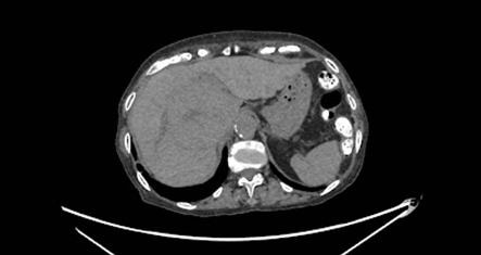
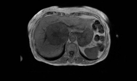
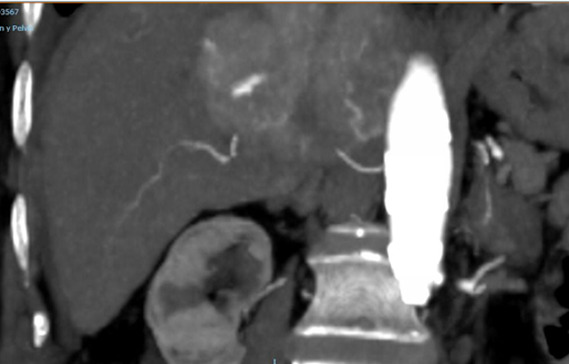
During an abdominal ultrasound ordered as part of her nephrology follow-up, a large solid hepatic mass was incidentally detected. The workup was completed with an abdominal CT scan and contrast-enhanced liver MRI, which revealed a lesion measuring approximately 8 cm located in segment VII, hypervascular in the arterial phase with washout in the late phases, consistent with hepatocellular carcinoma (LI-RADS 5), in a liver of normal size, contour, and parenchyma.
The case was presented to a multidisciplinary liver tumor committee. Given the partially atypical radiological appearance of the lesion and the absence of cirrhosis or known chronic liver disease, it was decided to perform a liver biopsy for diagnostic confirmation. The procedure, performed by the vascular radiology team, was complicated by the development of a perihepatic hematoma with active extravasation, which required a transfusion of packed red blood cells and subsequent conservative management.
The histopathological examination confirmed the diagnosis of hepatocellular carcinoma. Following a committee review and considering the patient’s age, functional status, and associated comorbidities, curative treatment options such as resection or liver transplantation were ruled out. Likewise, TACE and TARE were considered non-curative locoregional alternatives that were not appropriate in this case; therefore, systemic treatment with sorafenib was initiated.
Figure 1
Liver of normal size, contour, and echogenicity. A solid mass measuring approximately 8 × 6 cm is identified in the right hepatic lobe, located in segment VII, extending beyond the liver margin.
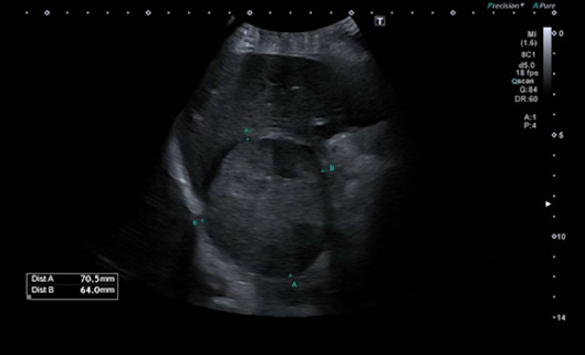
Figure 2
Liver of normal size and attenuation. A large, well-defined hepatic mass with central scarring is observed in segment VII, measuring approximately 7.5 × 8 × 7.7 cm.
Discussion
AIP is a rare condition with autosomal dominant inheritance and low penetrance, caused by a deficiency of porphobilinogen deaminase. Although it has traditionally been recognized for its acute neurovisceral crises, it is now known to be a multisystemic disease with significant chronic complications, including hypertension, chronic kidney disease, and liver neoplasms[1],[2]. This longitudinal perspective is important, as some patients may present with minimal acute clinical activity yet remain at risk for long-term complications.
From an epidemiological standpoint, the accumulated evidence shows a clear increase in the risk of hepatocellular carcinoma in acute hepatic porphyrias, especially in AIP. In a Swedish cohort of 1,244 patients with acute hepatic porphyria, the risk of primary liver cancer was significantly higher than in the general population, and in patients over 50 years of age with a history of biochemical activity, the annual incidence reached 1.8%[3]. Consistent with this, U.S. studies and a recent meta-analysis confirm that HCC can occur in the absence of cirrhosis and that the risk increases with age, particularly after age 50[4],[5].
This behavior distinguishes HCC associated with AIP from that observed in most cases of hepatocellular carcinoma in the general population, which are typically linked to advanced chronic liver disease or cirrhosis. Therefore, conventional screening programs based exclusively on cirrhosis may fail to identify these at-risk patients. Consequently, various authors and reference documents recommend periodic liver monitoring in patients with acute hepatic porphyria starting at age 50, generally through annual ultrasound, with or without alpha-fetoprotein testing according to local protocols[1]-[5].
The case presented here highlights how a lack of gastrointestinal follow-up can delay the diagnosis of a serious complication and limit available treatment options. The incidental detection of the tumor during an ultrasound examination ordered for another reason underscores the need for structured, protocol-based surveillance in patients with AIP. When the diagnosis is made at advanced stages or in elderly patients with comorbidities, potentially curative options may not be feasible; furthermore, locoregional treatments such as TACE or TARE, although useful in selected scenarios, should not be considered curative treatments in and of themselves.
With regard to the specific treatment of porphyria, the development of givosiran has represented a significant advance for patients with acute hepatic porphyria and recurrent attacks, as it has led to a sustained reduction in the annualized attack rate, the use of hemin, and the burden of symptoms[6]. However, its primary role is to control the underlying disease, and it does not replace liver monitoring, as the risk of liver cancer persists as a long-term complication that requires specific follow-up. In selected patients with severe AIP, liver transplantation may also correct the underlying metabolic defect, although its indication must be determined on a case-by-case basis according to age, comorbidities, and oncological status[1],[2].
From a practical standpoint, this case reinforces the need to establish specific follow-up programs for patients with AIP, coordinated among primary care, gastroenterology/hepatology, and other relevant specialties. Such programs should include education on precipitating factors, monitoring of chronic complications, and periodic liver imaging starting at age 50, even in the absence of cirrhosis or abnormal liver function tests. Early identification of liver lesions can broaden therapeutic options and decisively influence the clinical course.