
 Descargar número completo
 Download full issue
CITE THIS WORK
Menacho Ordóñez S, García Martínez A, Marín Lara L. Plummer-Vinson syndrome as an uncommon cause of progressive dysphagia.RAPD 2026;49(4):142-143. DOI: 10.37352/2026494.4
Introduction
Plummer-Vinson syndrome is a very rare clinical condition defined by the triad of iron-deficiency anemia, dysphagia, and esophageal membranes, and its presentation has been described predominantly in middle-aged women1. Its pathophysiology is not fully understood, although it has been proposed that iron deficiency promotes epithelial atrophy and impairments in mucosal repair2.
Clinical case
A 65-year-old woman was referred for progressive dysphagia with solids that had been present for 12 months, associated with weight loss. The symptoms were initially attributed to poorly fitting dentures and possible oral candidiasis, but there was no improvement following treatment.
Physical examination revealed glossitis, angular cheilitis, and a depapillated tongue. Laboratory tests showed iron-deficiency anemia.
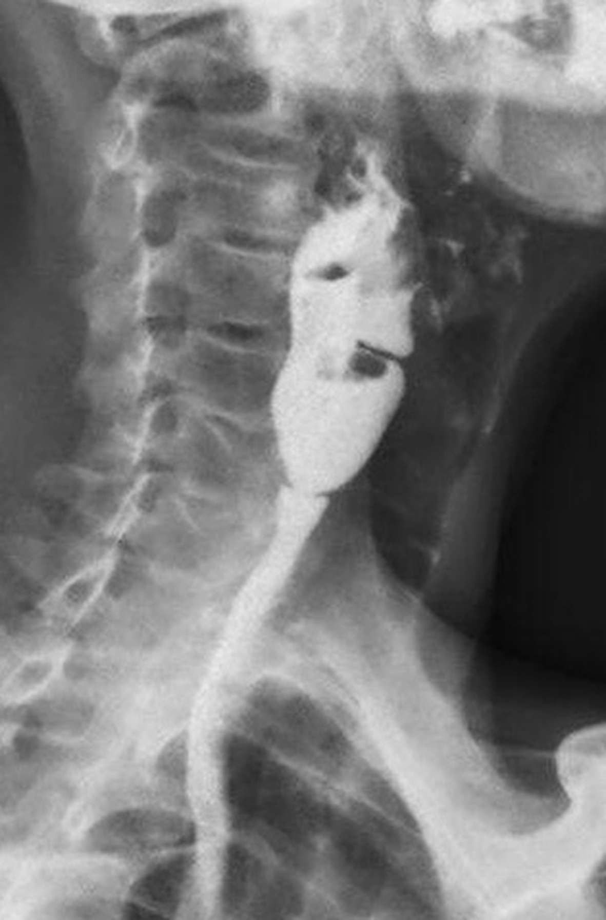
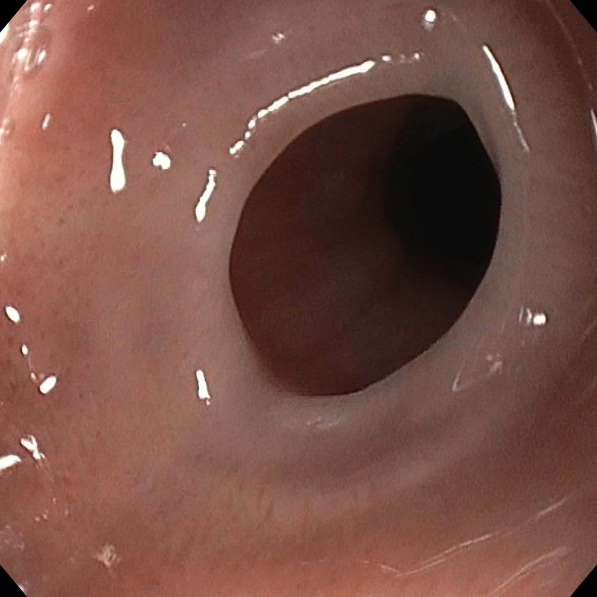
The esophagogram revealed a proximal esophageal membrane. Upper gastrointestinal endoscopy confirmed stenosis secondary to two cervical esophageal membranes without associated malignant lesions. Progressive endoscopic balloon dilation was performed (initially 7-;8 mm, increased to 10-12 mm in a second session) without complications, resulting in immediate symptomatic improvement. Treatment with oral iron was initiated, and laboratory values subsequently returned to normal.
Discussion
PVS is currently a rare cause of dysphagia in developed countries1. Its diagnosis may be delayed because symptoms are initially attributed to more common causes. Recognition of mucocutaneous signs of iron deficiency can guide the diagnosis.
Iron supplementation forms the basis of treatment and can improve dysphagia in the early stages, whereas in cases with established stenosis, endoscopic dilation is safe and effective1,2. Despite its current low prevalence in developed countries, it remains clinically significant due to its association with squamous cell carcinoma of the hypopharynx and esophagus, underscoring the importance of early diagnosis and regular clinical follow-up3.