
 Descargar número completo
 Download full issue
CITE THIS WORK
Pérez Sáez C, Fernández Carrasco M, Villegas Pelegrina P. Biliary hamartomatosis: the diagnostic challenge behind an incidental finding. RAPD 2026;49(4):147-149. DOI: 10.37352/2026494.6
Introduction
Multiple biliary hamartomas, also known as Von Meyenburg complex, are rare benign malformations that originate from the embryonic ductal plate.[1]. They are usually diagnosed incidentally on imaging studies, as most patients remain asymptomatic[2].
In some cases, they may cause elevated transaminases, pruritus, or be associated with chronic liver disease, and may lead to portal hypertension. Additionally, an association with hepatic and renal polycystic disease has been described[2].
The main differential diagnosis is multiple small liver metastases. The prognosis, however, is usually favorable.[2],[3].
Clinical case
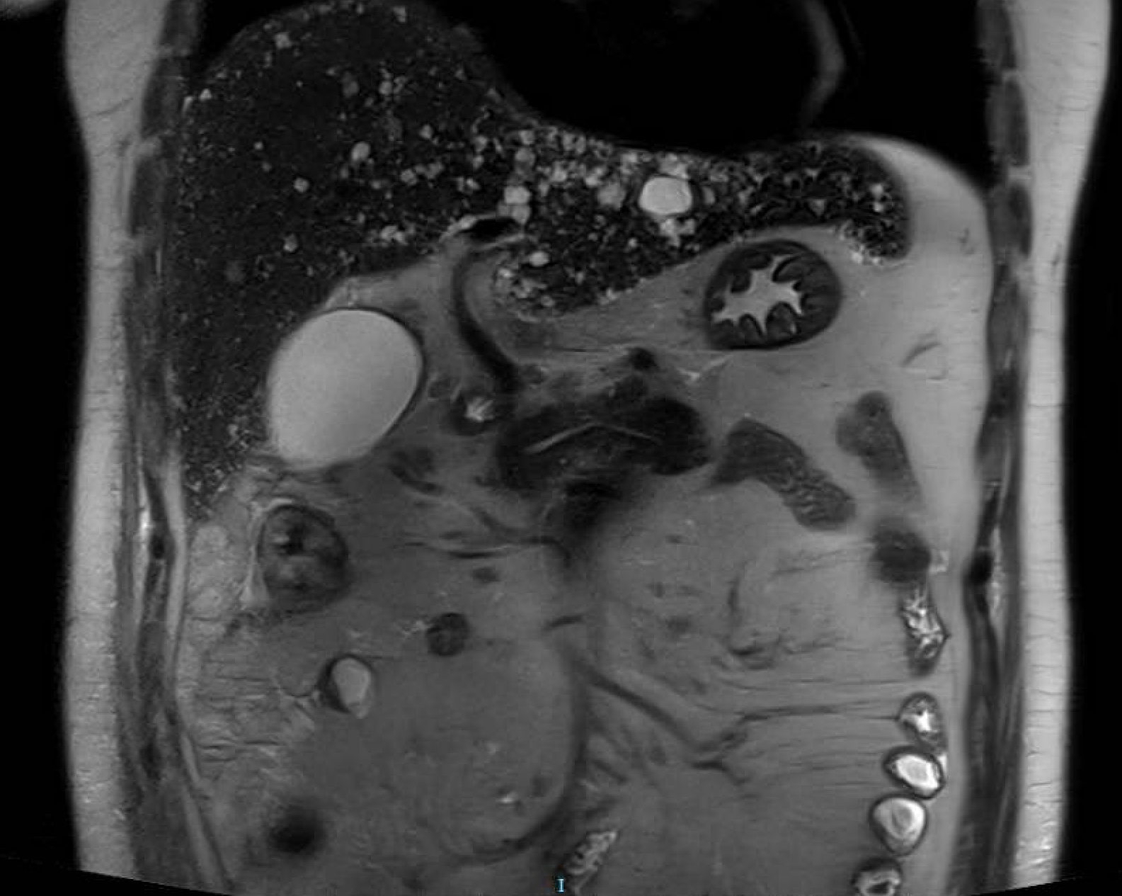
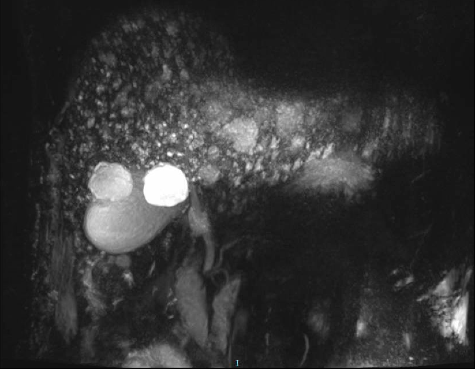
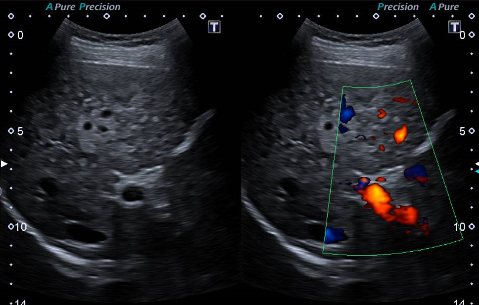
A 62-year-old woman with a personal history of hypothyroidism and hypercholesterolemia, occasional alcohol consumption, and under follow-up since 2020 for hypertransaminasemia with a cholestatic profile and pruritus. Chronic liver disease with significant fibrosis was documented (Fibroscan: 18.8 kPa). During the etiological workup, multiple subcentimeter-sized hepatic FLL were identified that were hyper-enhancing in the arterial phase and exhibited nonspecific behavior, with no apparent radiological signs of malignancy; additionally, the CA 19-9 marker level has been around 200 U/mL since the start of the workup. Finally, a liver biopsy was performed, revealing histological findings consistent with multiple biliary hamartomas associated with nodular regenerative liver changes.
Discussion
Biliary hamartomas can be located in any segment of the liver, although they tend to occur more frequently in the peripheral regions. They are usually small, measuring less than 1.5 cm in most cases[1],[2]. In their evaluation, the differential diagnosis must be considered against multiple cystic lesions, small, diffusely distributed metastases, infiltrative-pattern hepatocellular carcinoma, disseminated microabscesses, and multiple nodular steatosis.[2],[3]. On computed tomography, they appear as hypodense areas. On gadolinium-enhanced magnetic resonance imaging, they are characterized by T1-hypointense and T2-hyperintense signals.[1],[2].
The CA 19-9 tumor marker is nonspecific and may remain persistently elevated in patients with hamartomatosis without indicating malignancy. However, exceptional cases of transformation into intrahepatic cholangiocarcinoma have been described in the literature, particularly in patients with underlying chronic liver disease[3], which warrants individualized clinical follow-up in certain patients.
In general, biliary hamartomatosis does not require specific treatment, except in symptomatic patients (e.g., right upper quadrant pain or pruritus) or in those in whom a complication is suspected. The prognosis is usually favorable, although it is prudent to maintain clinical surveillance in patients with associated advanced liver disease[3].