
 Descargar número completo
 Download full issue
CITA ESTE TRABAJO
Plaza Fernández A, Moreno Moraleda I, Pérez Campos E. Green apple and red congo: atypical dysphagia colours. RAPD 2024;47(2):89-90. DOI: 10.37352/2024472.7
Clinical Case
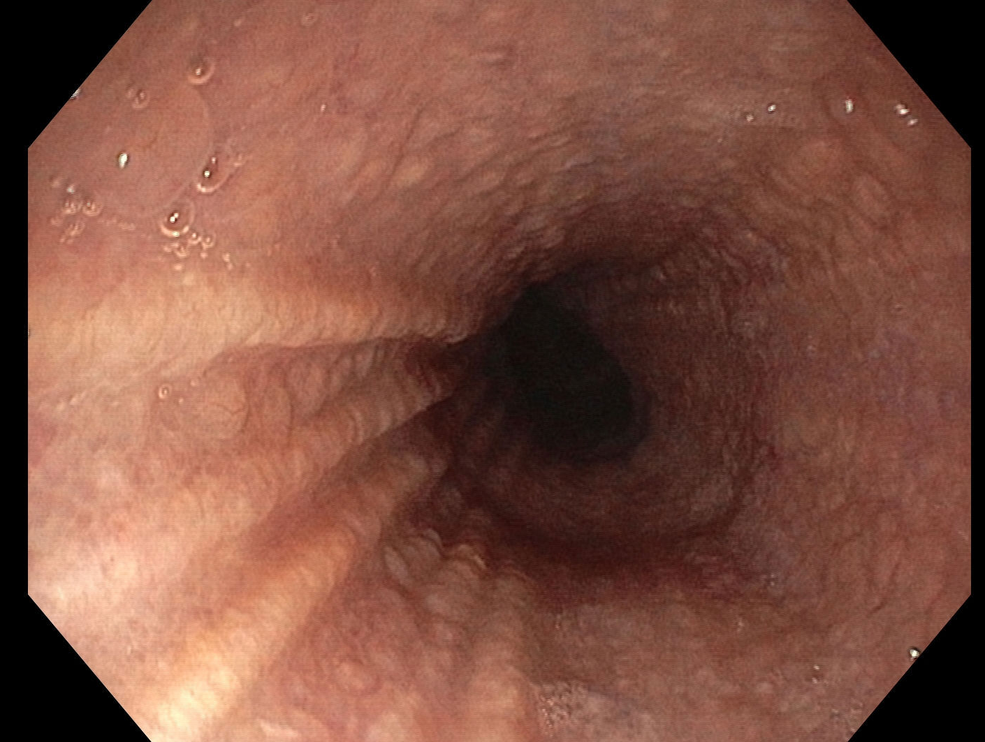
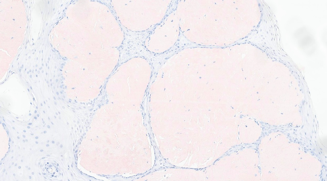
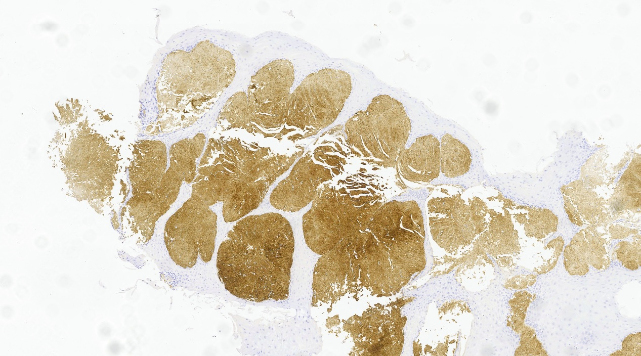
71-year-old male, hypertensive and dyslipidaemic. At the age of 40, he was referred to the otorhinolaryngology department for his debut with dyspnoea, and was diagnosed with laryngeal amyloidosis. At 70 years of age, he began with progressive dysphagia to solids and liquids, denying pyrosis, weight loss, abdominal discomfort or other symptoms, with unremarkable blood tests. Oesophagogastroduodenoscopy was performed, showing granular oesophageal mucosa (Figure 1), from which biopsies were taken. Barium swallow was also performed, which showed no evidence of swallowing disorder. The biopsies showed submucosal deposits of amorphous eosinophils, positive for Congo red staining (Figure 2), and serum amyloid P, with green birefringence by polarised light microscopy (Figure 3), these findings being compatible with the diagnosis of oesophageal amyloidosis.
Discussion
Oesophageal amyloidosis is usually silent and, if symptomatic, gastro-oesophageal reflux is the most common clinical manifestation. Dysphagia, on the other hand, is a rare entity in this context. Endoscopically, mucosal friability, erosions, ulcers and submucosal haematomas are usually observed. Given the variability of endoscopic findings, as well as the presentation of non-specific symptoms, confirmatory diagnosis requires histopathological studies such as Congo red staining or birefringence under polarised light.
The mechanism of dysphagia secondary to oesophageal amyloidosis is unknown, although part of it is attributed to a certain component of dysmotility secondary to atrophy due to nerve damage and pressure generated by the amyloid deposit as it settles between the muscle fibres. For this reason, the use of functional tests such as barium transit or oesophageal manometry are also of interest, as they can offer characteristic radiological patterns in some cases (oesophageal dilatation with distal narrowing).
To date, there are hardly any described cases of dysphagia as the main manifestation of this disease, so examples such as this one should help us not to forget this diagnostic possibility, integrating oesophageal amyloidosis into the differential diagnoses of dysphagia.