
 Descargar número completo
 Download full issue
CITA ESTE TRABAJO
Fernández Carrasco M, Rodríguez Mateu A, Villegas Pelegrina P. Giant epidermoid splenic cyst as a rare incidental finding. RAPD 2025;48(3):117-118. DOI: 10.37352/2025483.6
Clinical case
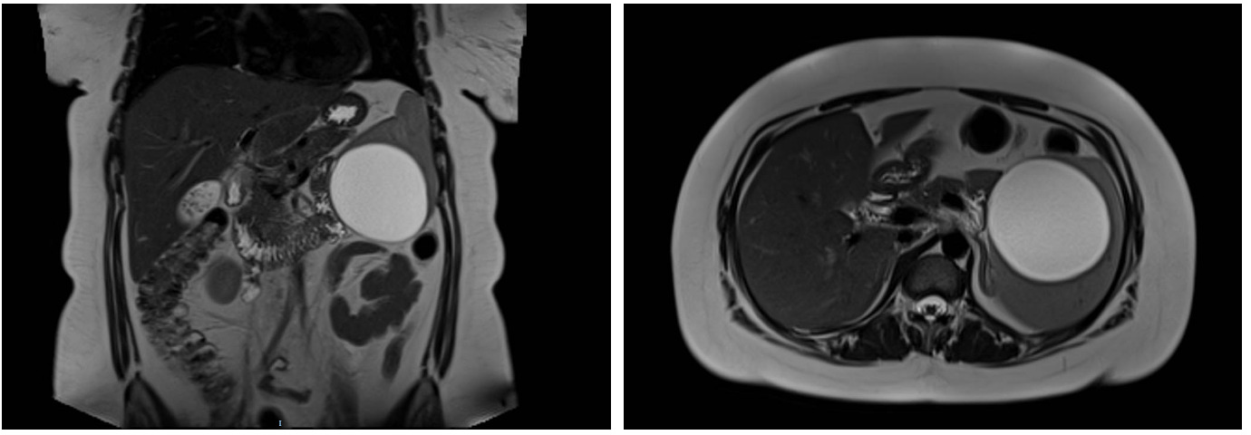
A 28-year-old female patient, with no relevant history, was admitted for complicated biliary colic. Laboratory tests showed elevated cholestasis enzymes (total bilirubin 3.7 mg/dL) and mild hypertransaminasemia (AST 70 U/L, ALT 91 U/L), without inflammatory markers. Abdominal ultrasound and magnetic resonance imaging (MRI) were performed with findings of multiple cholelithiasis without signs of cholecystitis and mild dilatation of the biliary tract without obstruction, incidentally revealing a 9 cm unilocular splenic cyst homogeneously hyperintense in T2 and hypointense in T1, without signs of complication.
Due to its large size, given the risk of complication, laparoscopic cystectomy was performed after preventive vaccination. The anatomopathological study confirmed a fibrous wall cyst lined by benign squamous epithelium, without malignancy.
Discussion
Primary splenic cysts are rare lesions, classified as primary (true) or secondary (pseudocysts). The primary ones include the epidermoid ones and are very rare, derived from embryologic anomalies. Secondary ones are usually post-traumatic, hemorrhagic, infectious or post-splenic infarction[1].
They are usually asymptomatic, being an incidental finding, but may present complications such as infection, hemorrhage or rupture. Their diagnosis is based on imaging studies. In ultrasound, epidermoid cysts appear as well-defined, thin-walled lesions with scattered internal echoes, and may present septa and trabeculations. On computed tomography (CT), these cysts appear as round, well-defined structures, without enhancement and with a water-like attenuation. On the other hand, false cysts, which lack a cellular lining, present more variable appearances on ultrasound, such as peripheral "eggshell" shaped calcifications and thick fibrous walls. On magnetic resonance imaging they appear hyperintense on T2 and hypointense on T1. Definitive confirmation requires histological analysis, showing the epithelial lining of the cyst[2].
Treatment of a splenic cyst may include total splenectomy, either open or laparoscopic. Options such as percutaneous drainage, sclerosis or partial splenectomy minimize recurrence and favor recovery[3]. Laparoscopic cystectomy is preferable in order to preserve splenic function and avoid the risk of receiving, which was the management performed in this patient[1],[2].