
 Descargar número completo
 Download full issue
Cite this work
Plaza Fernández A, Rodríguez Mateu A, Fernández Carrasco M. Liver amyloidosis as an exceptional cause of intrahepatic cholestasis. RAPD 2025;48(4):144-145. DOI: 10.37352/2025484.5
Introduction
Amyloidosis is a rare disease characterized by the extracellular deposition of insoluble proteins in the form of fibrils, which are resistant to proteolytic degradation. It can affect virtually any organ, causing progressive deterioration of its function. Hepatic involvement, although described, is rare and usually asymptomatic or with nonspecific symptoms. The presence of intrahepatic cholestasis and jaundice as an initial manifestation is exceptional. Below, we present a representative case of this unusual form of onset.
Clinical case
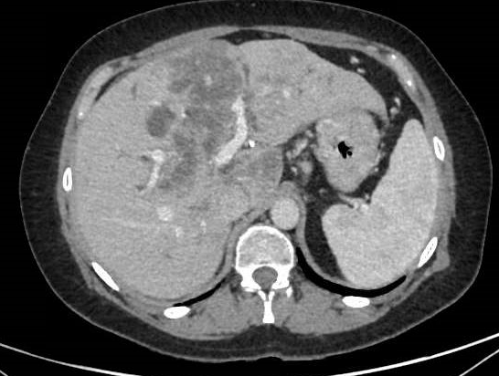
A 38-year-old patient with a history of obesity and type 2 diabetes mellitus presented to the emergency department with painless jaundice that had been present for one week. He reported progressive asthenia and weight loss of 5 kg in the last month. Laboratory tests showed total bilirubin of 8.1 mg/dL (direct 6.7 mg/dL), GGT 820 U/L, FA 1450 U/L, ALT 88 U/L, AST 72 U/L, and albumin 3.1 g/dL, with no significant elevation of tumor markers (CA 19.9, CEA, AFP). Abdominal ultrasound showed hepatomegaly, heterogeneous parenchyma with poorly defined hypoechoic areas, hyperechoic images suggestive of calcifications, and splenomegaly. The study was completed with computed tomography (CT), which showed multiple patchy hypodense areas in virtually all hepatic segments, some confluent, dilation of the intrahepatic bile duct, decreased intrahepatic vascular caliber, subcentimeter retroperitoneal adenopathies, and free fluid in the pelvis, initially pointing to intraductal cholangiocarcinoma (Figure 1). Given these findings, a liver biopsy was performed, which showed amyloid deposits positive with Congo red staining, confirming the diagnosis of hepatic amyloidosis.
Discussion
Hepatic amyloidosis is a rare manifestation of systemic amyloid disease, usually associated with protein deposits in the hepatic sinusoids, portal space, or bile ducts. It usually presents asymptomatically or with vague symptoms such as asthenia, mild elevation of liver enzymes, or, less frequently, jaundice and intrahepatic cholestasis[1],[2].
Imaging tests may show diffuse and heterogeneous liver involvement reflecting progressive liver damage due to amyloid infiltration, such as hypodense or hypocaptant areas that may mimic malignant lesions[3]. Liver biopsy with Congo red staining is the gold standard for diagnosis, allowing amyloid infiltration to be differentiated from other entities such as cholangiocarcinoma or lymphoma[4],[5].
In this context, given the variety of clinical manifestations of hepatic amyloidosis, it is essential to maintain a high index of diagnostic suspicion, especially in patients with nonspecific symptoms and ultrasound findings suggestive of diffuse liver involvement.