
 Descargar número completo
 Download full issue
CITE THIS WORK
Plaza Fernández A, Fernández Carrasco M, Rodríguez Mateu A. Beyond helicobacter pylori: gastric t-cell lymphoma as an unusual cause of gastrointestinal bleeding. RAPD 2025;48(6):215-216. DOI: 10.37352/2025486.2
Introduction
Primary gastric lymphomas represent a small percentage of gastric neoplasms, and among them, B-cell lymphomas, such as MALT lymphoma and diffuse large B-cell lymphoma, are the most common. In contrast, primary gastric T-cell lymphoma is extremely rare and poses a diagnostic challenge due to its nonspecific clinical presentation. We present a clinical case that presented initially as gastrointestinal bleeding.
Clinical case
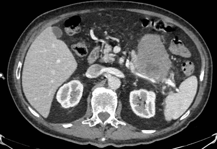
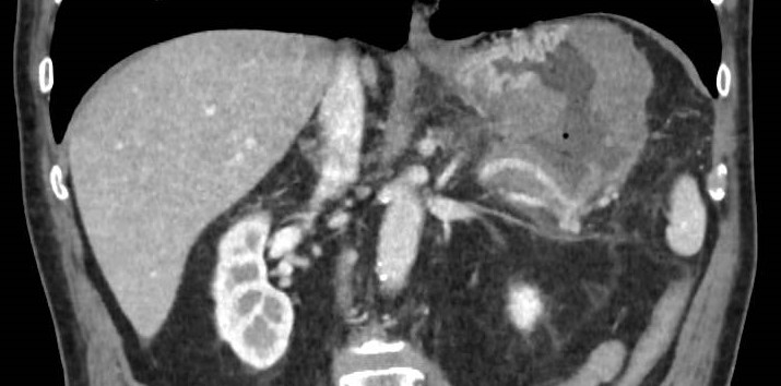
A 75-year-old patient with a history of Helicobacter pylori infection eradicated years ago and resected urothelial carcinoma currently in remission. The patient had no known autoimmune diseases or viral infections. In the months prior to admission, the patient reported progressive asthenia and weight loss of approximately 6 kg, with no other relevant digestive symptoms. He went to the emergency department due to an episode of hematemesis associated with syncope. Laboratory tests revealed anemia in the transfusion range, and an urgent gastroscopy was performed, which revealed a large ulcerated infiltrative mass in the gastric body at the level of the greater curvature, with irregular and friable edges and active bleeding. Hemostatic control was achieved by sclerotherapy with adrenaline and the application of hemostatic powders. Subsequently, the patient presented a new episode of hematemesis with hemodynamic instability, so a computed tomography (CT) angiogram was performed, which revealed active bleeding from the gastric lesion. Given the instability, urgent surgery was indicated, and a partial gastrectomy was performed with control of the hemorrhagic focus using hemostatic sutures. Subsequently, during admission, review of the previous CT images revealed infiltration of the tail of the pancreas and splenic artery, as well as perigastric and retroperitoneal lymphadenopathy, with no distant involvement (Figures 1 and 2). Histological analysis showed diffuse high-grade T-cell lymphoid proliferation, with positivity for CD3 and CD7 and negativity for B markers, confirming the diagnosis of gastric T-cell lymphoma. The patient was referred to Hematology and started treatment with systemic chemotherapy (modified CHOP). The initial course was favorable, with a good clinical response and no new bleeding episodes during short-term follow-up.
Discussion
Primary gastric T-cell lymphoma is an extremely rare neoplasm, with a very low incidence compared to B-cell lymphomas, among which MALT lymphoma is the most common subtype[1],[2]. Unlike these, its association with Helicobacter pylori is not well established, although it has been linked to viral infections such as HTLV-1, HBV, HCV, or HIV, and to autoimmune diseases[2],[3].
The clinical presentation is usually nonspecific, with symptoms such as abdominal pain, weight loss, and asthenia predominating. Gastrointestinal bleeding, as in our case, is an unusual form of onset[4]. Endoscopically, these lymphomas can take many forms: infiltrating masses, ulcers, or thickening of folds[5].
Diagnosis requires biopsy with immunohistochemical study, with the CD3+ and CD20− phenotype being characteristic. Echoendoscopy allows assessment of transmural involvement and locoregional lymphadenopathy, while CT is essential for assessing distant disease. Treatment is usually based on chemotherapy, with regimens such as CHOP, although the course is often aggressive and the prognosis is poor[2],[4].
In conclusion, primary gastric T-cell lymphoma is a rare entity with a complex diagnosis. Its atypical presentation, such as gastrointestinal bleeding, requires a high index of suspicion for timely diagnosis and treatment.