
 Descargar número completo
 Download full issue
CORRESPONDENCE
Gloria Francisca León Sanjuan
Jerez Hospital. Jerez de la Frontera, Cádiz.
11407 Jerez de la Frontera
CITE THIS WORK
León Sanjuan GF, García Martínez A, Benavente Oyega MA. Esophagitis dissecans superficialis in an HIV patient with a history of substance abuse. RAPD 2025;48(6):217-218. DOI: 10.37352/2025486.3
Introduction
Esophagitis dissecans superficialis (EDS) is characterised by circumferential desquamation of the oesophageal mucosa, causing striking lesions that usually have a benign course. We present the case of a patient with HIV infection and substance abuse.
Clinical case
A 58-year-old male, daily user of cannabis, cocaine, and inhaled heroin. Diagnosed in 2004 with human immunodeficiency virus (HIV) infection, currently stage C3, with regular adherence to treatment for years. History of hepatitis C virus infection, treated and cured with direct-acting antivirals.
He was admitted to the Infectious Diseases Unit for asthenia, pneumococcal pneumonia, oropharyngeal candidiasis, and cachexia. In this context, a chest CT scan was performed, showing evidence of thickening of the oesophageal wall in the distal third. Therefore, an oral endoscopy was requested, which identified a circumferential area of desquamative mucosa with friability on rubbing, compatible with oesophagitis, approximately 32 cm from the dental arch (Figure 1).
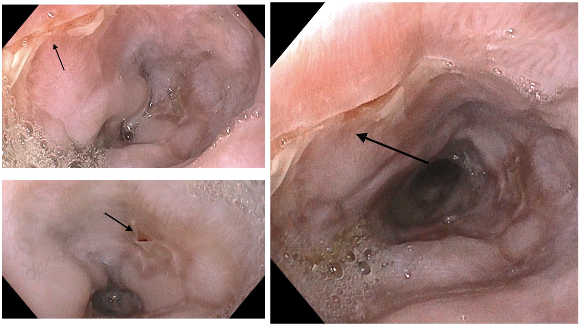
Figure 1
. Oesophageal endoscopic image showing areas of desquamated oesophageal mucosa, with the appearance of a partially detached membrane, without ulceration or active bleeding, findings consistent with esophagitis dissecans superficialis.
Biopsies were taken from the surrounding mucosa for microbiology and anatomopathological study, with no evidence of malignancy or viral induction, as well as negative immunohistochemistry for cytomegalovirus. No other pathological findings were evident on endoscopy.
Given the clinical context, exposure to multiple toxins, and the endoscopic and histological data, a compatible diagnosis was established: Esophagitis dissecans superficialis probably related to damage to the oesophageal mucosa caused by toxins.
Discussion
Superficial dissecting oesophagitis is a rare and often underdiagnosed condition. Its diagnosis is based on the finding of characteristic endoscopic lesions such as diffuse mucosal desquamation and the presence of ‘whitish membranes’, given that it can often be asymptomatic and histological examination is not specific in this condition[1],[2].
Although the available literature on this subject is limited, this condition has been associated with factors such as physical and chemical trauma, smoking, autoimmune bullous dermatoses, consumption of toxic substances, and certain drugs such as bisphosphonates, NSAIDs, doxycycline, and apixaban.
Despite its striking endoscopic appearance, it has a benign course, achieving complete healing after removal of the causative agent, if it is a drug or toxin, or through control of the underlying disease[3].
In the case we present, the findings are consistent with esophagitis dissecans superficialis associated with the consumption of toxic substances such as cocaine and heroin, in the context of possible mucosal fragility due to HIV.