
 Descargar número completo
 Download full issue
CITE THIS WORK
Cano de la Cruz JD, Sánchez Sánchez MI, Diego Martínez R, Bravo Aranda AM. Type IVA choledochal cyst diagnosed in adulthood. RAPD 2025;48(6):225-226. DOI: 10.37352/2025486.6
Clinical Case
We present the case of a 33-year-old woman admitted for pain in the right hypochondrium and fever. Laboratory tests reveal abnormal liver function (cytolysis and dissociated cholestasis).
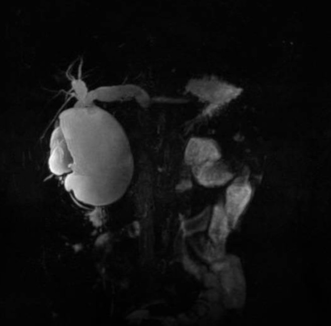
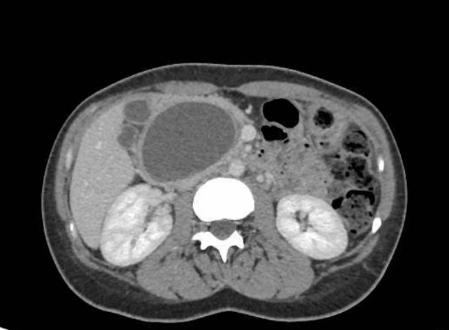
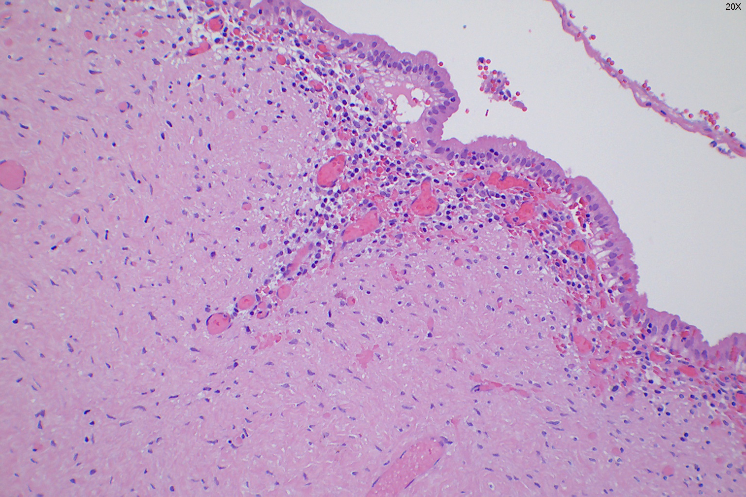
An abdominal ultrasound is performed, which shows dilation of the intrahepatic and extrahepatic bile ducts, with a common bile duct measuring up to 15 mm. An abdominal CT scan and MR cholangiography were performed, revealing marked cystic dilatation of the common bile duct (90 x 57 x 58 mm), associated with cystic dilatation of the intrahepatic bile duct, predominantly on the left side, findings suggestive of a type IVA common bile duct cyst (Figures 1-2). Clinical improvement was observed after administration of analgesia and empirical antibiotic therapy. It was decided to perform a cephalic duodenopancreatectomy (CDP) due to the intrapancreatic component of the lesion and the potential risk of malignancy, which was carried out without complications. After anatomopathological analysis of the surgical specimen (Figure 3), malignant degeneration of the lesion was ruled out.
Figure 1
MR cholangiography. Coronal section - Marked cystic dilatation of the common bile duct (90 x 57 x 58 mm) associated with cystic dilatation of the intrahepatic bile duct, predominantly on the left side.
Discussion
Choledochal cysts are rare entities consisting of a congenital dilation of the bile duct. They are mostly diagnosed in children, although in recent years there has been an increase in their incidence in the adult population. According to Todani's classification[1], the most common (80-90%) are type I (dilatation of the extrahepatic bile duct), while type IVA cysts involve dilatation of both the extrahepatic and intrahepatic bile ducts and are extremely rare (1-2%). In 70-80% of cases, they are related to an abnormal pancreatico-biliary junction (AUPJ), predisposing to reflux of pancreatic secretions, with proteolytic activity on the common bile duct[2].
In adults, they manifest as pain in the right hypochondrium, jaundice, pancreatitis, or cholangitis, with diagnosis primarily based on imaging tests, particularly MR cholangiography due to its high sensitivity (90-100%).
The treatment of choice will always be surgical, with complete removal of the cyst, given its potential risk of malignancy[3].